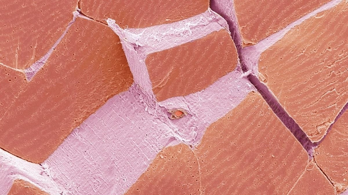
If you’ve ever stared at a microscopic image and questioned your life choices, you are not alone. This is the weekly Microscope Mystery. We drop a head-scratching visual into our newsletter and ask you to guess what it is. Sometimes the results are laughable.
Once, an assistant editor claimed a solar panel surface was a biscuit. Another journalist thought rose petal glands looked like burnt tastebuds. We are taking this feature off our newsletter shelves and onto the main site.
Introducing the Monday Microscope Mystery.
This week, we’re looking at a strange pink and purple landscape that looks dangerously edible. What do you think it is?
You can narrow your guesses by considering these four options:
- a. Ocean sponge
- b. Shark scales
- c. Skeletal muscle
- d. Lizard tongue
Don’t scroll past the spoiler yet. Watch orcas punch sunfish until they explode below. Then come back. You’ve been warned.
Decoding the Image: Skeletal Muscle
The answer is c. Skeletal muscle.
Look closely at those blocky, reddish-pink shapes. Each one is a single muscle fiber. It’s a cell so specialized it can stretch across the entire length of some of your body’s muscles.
Stack a few hundred fibers, and you get a fascicle. Stack a few hundred fascicles, and you get the bicep you use to lift groceries or the hamstring you use to walk.
The light purple webbing weaving through these fibers is called perimysium. It’s not just packing material. It’s filled with blood vessels and nerve endings. It wraps every fiber to keep them contracting in sync. Without it, the tissue would shear apart.
“Each of those blocky reddish-pinka shapes is a single muscle fiber… Bundle a few hundred fascicles together and you’re looking at a bicep or hamstring.”
Find the tiny, textured oval peeking out from the pink band near the center? That is a capillary. One of thousands of microscopic tubes threading through your muscle. They deliver oxygen the moment you move. Without them, those fibers would burn out.
False Colors and High Magnification
Here’s the catch: none of that color is real.
We captured this using a scanning electron microscope (SEM). This tool fires electrons at the sample instead of photons. Light microscopes can’t handle the detail. The resulting image is naturally black and white.
The reds and pinks? Added in post. Sometimes for aesthetics. Mostly so your eye can instantly distinguish muscle tissue from blood vessels.
The magnification here is 3,000x, assuming the final print is 10 cm wide.
What makes this tissue remarkable isn’t just the structure. It’s the regeneration. Skeletal muscle relies on satellite cells to repair damage. But those cells stay primed only if you move. Sit too long, and they lose the ability to divide. Dormancy kills the repair mechanism.
So when you shiver to stay warm, or flex your arm, remember: you are operating a self-repairing, wired, tiled machine.
It takes thousands of fibers working in unison to hold your body together. At this magnification, it just looks like a cracked tile floor waiting to happen.